カレントテラピー 32-7 サンプル page 17/32

このページは カレントテラピー 32-7 サンプル の電子ブックに掲載されている17ページの概要です。
秒後に電子ブックの対象ページへ移動します。
「電子ブックを開く」をクリックすると今すぐ対象ページへ移動します。
概要:
カレントテラピー 32-7 サンプル
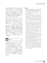
Current Therapy 2014 Vol.32 No.7 25狭心症の診断と治療641投与および冠動脈内投与で使用されるが,気管支喘息患者に禁忌であり,パパベリンは冠動脈内投与が行われるが,QT延長をきたすため,まれに心室細動が出現することがあり注意を要する.心筋血流全体を評価する心筋部分血流予備量(FFRmyo)は平均中心静脈圧(Pv),狭窄遠位部の平均冠動脈圧(Pd)および冠動脈狭窄近位部の平均冠動脈圧(Pa)を測定し,下記式により求められる10).FFRmyo=(Pd-Pv)/(Pa-Pv)(Pvが高くなければFFRmyoはPd/Paにて近似される)FFRmyo<0.75が機能的に有意であり,高い診断感度(90%),特異度(93%)を示すといわれており,自転車エルゴメータ負荷試験,エルゴメータ負荷タリウム心筋血流イメージング,ドブタミン負荷心エコー図法などの負荷試験の陽性率と強い相関を示す(図7)10).FFRは再現性に優れており,冠動脈狭窄の機能的重症度を評価できるため,冠動脈造影上中等度の狭窄で冠動脈造影所見だけではその狭窄が有意であるか否かの判断が困難な症例において,非常に有用な生理学的評価方法となる.冠動脈血流をNavier -Stokes方程式を用いてコンピュータ解析することによって,FFRを評価する(FFRCT)非侵襲的な方法も報告されている(図8)11).Ⅲ おわりに診断の役割として重視されるべきことは,治療方針の決定および予後に対するインパクトであって,必ずしも新しい検査法が優れているとは限らない.新しい診断技術が日常臨床に組み込まれるためには,その新しい検査法が以前のものに勝り,かつ,費用対効果にも優れていることを,科学的エビデンスをもって示す必要がある.そのため狭心症の病態把握,診断を進めていくうえで,各検査法の特質,診断性能,どのような診断目的や病態解析に有効か,またどのようなピットホールがあるか,などを熟知する必要がある.参考文献1)循環器病の診断と治療に関するガイドライン(2007-2008年度合同研究班報告):冠動脈病変の非侵襲的診断法に関するガイドライン.Circ J 73(Suppl Ⅲ):1019-1089, 20092)Kwok JM, Miller TD, Hodge DO, et al:Prognostic value ofthe Duke treadmill score in the elderly. J Am Coll Cardiol39:1475-1481, 20023)Gibbons RJ, Balady GJ, Bricker JT, et al;American Collegeof Cardiology/American Heart Association Task Force onPractice Guidelines(Committee to Update the 1997 ExerciseTesting Guidelines):ACC/AHA 2002 guideline update forexercise testing:summary article:a report of the AmericanCollege of Cardiology/American Heart Association TaskForce on Practice Guidelines(Committee to Update the 1997Exercise Testing Guidelines). Circulation 106:1883-1892,20024)Pierard LA, De Landsheere CM, Berthe C, et al:Identificationof viable myocardium by echocardiography during dobutamineinfusion in patients with myocardial infarction afterthrombolytic therapy:comparison with positron emissiontomography. J Am Coll Cardiol 15:1021-1031, 19905)Previtali M, Poli A, Lanzarini L, et al:Dobutamine stressechocardiography for assessment of myocardial viability andischemia in acute myocardial infarction treated with thrombolysis.Am J Cardiol 72:124G-130G, 19936)Bansal M, Jeffriess L, Leano R, et al:Assessment of myocardialviability at dobutamine echocardiography by deformationanalysis using tissue velocity and speckle -tracking.JACC Cardiovasc Imaging 3:121-131, 20107)Hachamovitch R, Berman DS, Shaw LJ, et al:Incrementalprognostic value of myocardial perfusion single photon emissioncomputed tomography for the prediction of cardiacdeath:differential stratification for risk of cardiac death andmyocardial infarction. Circulation 97:535-543, 19988)Gaemperli O, Bengel FM, Kaufmann PA:Cardiac hybridimaging. Eur Heart J 32:2100-2108, 20119)Ishida N, Sakuma H, Motoyasu M, et al:Noninfarcted myocardium:correlation between dynamic first-pass contrastenhancedmyocardial MR imaging and quantitative coronaryangiography. Radiology 229:209-216, 200310)Pijls NH, De Bruyne B, Peels K, et al:Measurement of fractionalflow reserve to assess the functional severity of coronaryartery stenosis. N Engl J Med 334:1703-1708, 199611)Nakazato R, Park HB, Berman DS, et al:Noninvasive fractionalflow reserve derived from computed tomography angiographyfor coronary lesions of intermediate stenosis severity:results from the DeFACTO study. Circ CardiovascImaging 6:881-889, 2013